Home ![]() WHY MIGHT I BE CALLED BACK AFTER MY SCREENING MAMMOGRAM?
WHY MIGHT I BE CALLED BACK AFTER MY SCREENING MAMMOGRAM?
A screening mammogram uses specialized X-ray technology to take a series of images of the breasts and provide a detailed look at the internal structure of breast tissue. Sometimes after your screening mammogram, you may be asked to return for additional breast imaging, such as a diagnostic mammogram or breast ultrasound.
You would be called back for a diagnostic mammogram to examine a specific area of concern, to assess a change in the breast tissue compared to previous images, or to investigate an abnormality.
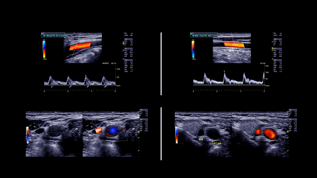
In addition to a diagnostic mammogram, you may also be sent for a breast ultrasound if it’s determined on a mammogram that you have high breast density. You could have either an automated or handheld breast ultrasound which use high frequency sound waves to see the internal structure of your breast. It can be requested to examine a specific area of concern that has been identified on a mammogram. Breast ultrasound helps provide a more detailed look and ensures you have received a complete breast exam.
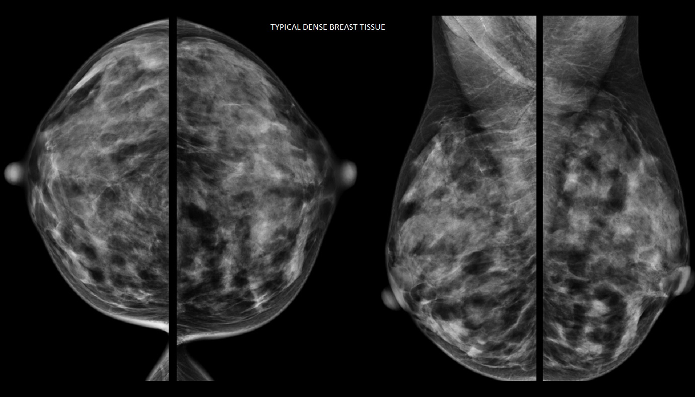
Dense breast tissue can only be determined by mammography and is not something that can be felt or seen. Dense breasts have less fat and more fibroglandular tissue, which can make it difficult to detect changes in the breast and small cancers may be hidden.
Dense breast tissue also increases the risk of breast cancer, especially in older women. For women with dense breast tissue in 75% or more of their breasts, their breast cancer risk is double relative to the general population. Using both mammography and ultrasound together can help provide a more complete picture of your dense breast tissue.
According to the Canadian Cancer Society, one in eight Canadian women will be affected by a breast cancer diagnosis in their lifetime. However, when breast cancer is detected early, before it’s clinically apparent (e.g., palpable lump), it’s more likely to be small and more easily treated. Small cancers detected early can be removed and breast conserving surgery can be performed. Additionally, small cancers often do not require chemotherapy or radiation therapy.
The best way to detect breast cancer early is through regular screening mammograms. Your Alberta Health Care Insurance Plan covers yearly mammograms starting at age 40. Having regular mammograms allows a radiologist to compare your images from year to year, identifying changes that may be occurring in the breast over time.
Mayfair Diagnostics follows the Canadian Association of Radiologists guidelines and recommends yearly mammograms from age 40-49. After that it’s recommended that women have a mammogram every two years from age 50-74. However, if you have a first degree relative (parent, sibling, child) with a family history of breast cancer or your own medical history indicates a higher risk of developing breast cancer, your doctor may recommend continuing to have a mammogram every year over age 50.
As part of the Alberta Breast Screening program, women 50-74 years of age do not need a referral from a doctor to have a screening mammogram. After age 75, screening frequency will depend on your medical history and should be discussed with your doctor. It is also important to be aware of any changes in your breasts, such as new pain or lump, nipple discharge, or any heat or redness, and to report those to your doctor.
The amount of radiation exposure from mammography is comparable to the exposure every person gets from the earth’s natural background radiation from living in Calgary over a period of six months. In most cases, the benefits of detecting breast cancer early with mammography outweighs the risk of exposure to this low dose of radiation.
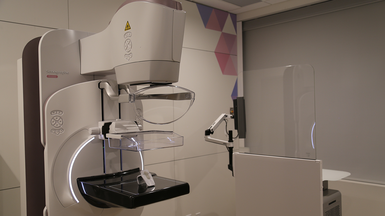
During a mammogram, two X-ray plates slowly move together, compressing the breast for a short amount of time. This compression is necessary to get the clearest images of your breast tissue using the least amount of radiation. Since individuals experience pain differently, there may be some discomfort during the very short interval when the breast tissue is being compressed, however it should be minimal.
Mayfair Diagnostics has 12 locations which offer mammography exams, and except for our Coventry Hills all of them use the Senographe Pristina mammography system – which helps provide a more comfortable mammogram. Visit our breast imaging page for more information.
Alberta Health Services (2021) “Breast Screening.” www.screeningforlife.ca. Accessed October 1, 2022.
Canadian Association of Radiologists (2016) “CAR Practice Guidelines and Technical Standards for Breast Imaging and Intervention.” www.car.ca. Accessed October 1, 2022.
Canadian Cancer Society (2018) “Breast cancer statistics.” www.cancer.ca. Accessed October 1, 2022.
Coldman, A., et al (2014) “Pan-Canadian Study of Mammography Screening and Mortality from Breast Cancer.” Journal of the National Cancer Institute. November 2014, 106 (11).
Monticciolo, Dr. et al. (2018) “Current Issues in the Overdiagnosis and Overtreatment of Breast Cancer.” American Journal of Roentgenology. February 2018, 210 (2). Accessed October 1, 2022.
Tabar, L., et al. (2019) “The incidence of fatal breast cancer measures the increased effectiveness of therapy in women participating in mammography screening.” Cancer. Accessed October 1, 2022.
© 2024 Mayfair. All rights reserved.
| Cookie | Duration | Description |
|---|---|---|
| cookielawinfo-checkbox-analytics | 11 months | This cookie is set by GDPR Cookie Consent plugin. The cookie is used to store the user consent for the cookies in the category "Analytics". |
| cookielawinfo-checkbox-functional | 11 months | The cookie is set by GDPR cookie consent to record the user consent for the cookies in the category "Functional". |
| cookielawinfo-checkbox-necessary | 11 months | This cookie is set by GDPR Cookie Consent plugin. The cookies is used to store the user consent for the cookies in the category "Necessary". |
| cookielawinfo-checkbox-others | 11 months | This cookie is set by GDPR Cookie Consent plugin. The cookie is used to store the user consent for the cookies in the category "Other. |
| cookielawinfo-checkbox-performance | 11 months | This cookie is set by GDPR Cookie Consent plugin. The cookie is used to store the user consent for the cookies in the category "Performance". |
| viewed_cookie_policy | 11 months | The cookie is set by the GDPR Cookie Consent plugin and is used to store whether or not user has consented to the use of cookies. It does not store any personal data. |