
Home ![]() WHAT DOES COMPUTED TOMOGRAPHY MEAN?
WHAT DOES COMPUTED TOMOGRAPHY MEAN?
Computed tomography scans are often referred to as CT or CAT scans. It’s a type of medical imaging that uses ionizing electromagnetic radiation in the form of X-ray photons (energy) to create images of your body.
Conventional X-ray imaging uses a fixed X-ray source, which emits photons that travel through the body part of interest. An X-ray detector is placed behind the area of interest to record the signal of the photons after their journey, resulting in a two-dimensional image.
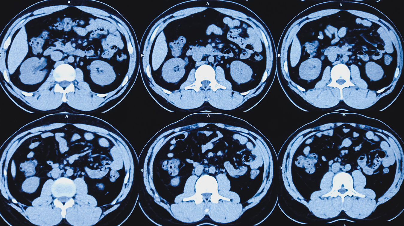
During a CT scan, a rotating X-ray source emits a narrow beam of photons through the body part of interest which is then recorded by an advanced detector on the other side. This results in a volumetric dataset that the computer reconstructs into numerous cross-sectional images, called tomographic images or slices. This creates three-dimensional (3D) images that can be viewed from multiple directions.
Since its invention by Godfrey N. Hounsfield at Electric and Musical Industries (EMI) in 1971, CT has proven to be a sophisticated type of medical imaging that provides detailed anatomic images of the body. It also has the ability to evaluate the arteries, veins, and blood flow to organs when augmented with intravenous contrast.
CT scans are very good at imaging bone but can also provide detailed information about soft tissues in the chest, abdomen, and pelvis, in addition to blood vessels. A CT may be preferred to evaluate fractures or look for cancers or blood clots.
CT lung screening utilizes a lower radiation dose technique that can help to determine the presence of, and then further evaluate, suspicious lung nodules for lung cancer, as well as other serious illnesses. It could be appropriate for patients at high risk of lung cancer due to smoking, exposure to radon gas, a family or personal history of lung cancer, or other risk factors. In these cases, CT lung screening can help detect early forms of lung cancer, as small as a few millimeters in size.
For those at risk of coronary artery disease, a coronary CT angiography can be used to non-invasively examine the coronary arteries. It can detect both calcified or hard plaques and noncalcified or soft plaques, both of which have been implicated in adverse cardiac events. The presence of soft plaques is often best determined by IV contrast enhanced CT angiography.
CT virtual colonscopy utilizes a lower radiation dose technique to screen the large intestine (colon) and rectum for colon cancer. This requires insufflation of the colon with CO2 gas to permit better visualization. This differs from a colonoscopy in that an endoscope is not used.
At Mayfair Diagnostics we provide CT lung screening, CT coronary angiography, and CT virtual colonoscopy as private pay examinations at our Mayfair Place location. They can be purchased for single or multiple body areas. We also offer Health Assessment packages, which provide a discount on multiple imaging exams when purchased together.
Your health spending account or group medical insurance plan may cover the cost of a private CT that is prescribed by a qualified health care practitioner. You will need to check with your plan administrator for coverage details.
Whether public or private, a CT must be requested by a health care practitioner. To determine whether a CT is recommended, you will need to discuss with your doctor your medical and family history, risk factors, and if there are symptoms, how long symptoms have been present and how they affect daily activities.
If a private CT scan is indicated as a best next course of action, a requisition will be provided, and the appointment can be booked. It’s important to note that the exposure to radiation from a CT scan is higher than that of a conventional X-ray, as it is a more advanced imaging modality, but the associated risk is still small. For example, the radiation exposure from one low-dose CT scan of the chest is less than the exposure from the earth’s natural background radiation over six months. In most cases, the benefits of a CT, such as the early detection of a serious illness, outweigh the potential health risk from receiving a higher radiation dose.
For more information, please visit our services page.
REFERENCES
Alberta Health Services (2022) “New screening program will use CT scans to detect early-stage lung cancer.” screeningforlife.ca/lung. Accessed August 26, 2024.
Canadian Cancer Society (2024) “Colorectal cancer statistics.” cancer.ca. Accessed August 26, 2024.
Canadian Cancer Society (2024) “Risk factors for colorectal cancer.” cancer.ca. Accessed August 26, 2024.
Diagnostic Imaging, Alberta Health Services (2021) “Computed tomography (CT).” myhealth.alberta.ca. Accessed August 26, 2024.
De Koning, H. J., et al. (2020) “Reduced Lung-Cancer Mortality with Volume CT Screening in a Randomized Trial.” New England Journal of Medicine, 2020; 382:503-513. Accessed August 26, 2024.
National Institute of Biomedical Imaging and Bioengineering (2022) “Computed Tomography (CT).” nibib.nik.gov. Accessed August 26, 2024.
Schulz, R. A., et al. (2021) “How CT happened: the early development of medical computed tomography.” Journal of Medical Imaging. 2021 Sep; 8(5): 052110. Accessed August 26, 2024.
Radiologists are specialized physicians who interpret diagnostic imaging to diagnose and monitor a wide range of medical conditions. At Mayfair Diagnostics, they review X-ray, ultrasound, CT, and MRI studies, among others, analyzinsg images in detail and providing comprehensive reports and clinical recommendations to referring physicians. They collaborate closely with technologists and clinic teams to guide imaging protocols, ensure quality and radiation safety standards, and may perform image-guided procedures such as biopsies or injections. Through their expertise and teamwork, radiologists play a key role in accurate diagnosis, effective treatment planning, and improved patient outcomes.
Administrative professionals support the organization across human resources, marketing, operations, strategic partnerships, finance, information technology, and infrastructure. From recruiting staff to promoting services and improving workflows, they help ensure smooth operations and positive experiences for employees and patients. Through collaboration with clinical and support teams, they provide essential coordination that enables efficient, high-quality service.
Diagnostic Imaging Assistant (DIA) support clinic operations and help ensure a positive patient experience. They assist staff by greeting and preparing patients, confirming information, coordinating appointments, and guiding patients through their visit. DIAs also maintain exam rooms, manage documentation, and ensure supplies and equipment are ready. Through strong customer service, attention to detail, and teamwork, they help create a safe and organized environment.
Patient Experience Coordinators (PECs) are the first point of contact, scheduling exams and ensuring accurate patient information. They communicate clearly with patients, coordinate with care teams, and support a smooth, confidential, and customer-focused experience.
Nuclear Medical Technologists perform diagnostic nuclear medicine procedures involving sensitive and highly personal patient circumstances. They are responsible for delivering the highest standard of care in a professional, compassionate, and patient-centered manner, in accordance with provincial regulatory requirements, CAMRT standards, and Mayfair policies and guidelines.
Computed Tomography (CT) Technologists operate CT imaging equipment to produce detailed cross-sectional images of the body that assist in diagnosing a wide range of medical conditions. They prepare and position patients for scans, ensure safety protocols are followed, and administer contrast agents when required. CT technologists work closely with radiologists to ensure high-quality diagnostic images are obtained, while providing clear communication and compassionate care to support patient comfort throughout the procedure.
Magnetic Resonance Imaging (MRI) Technologists operates MRI scanners to produce detailed images of internal body structures used to assist in medical diagnosis and treatment planning. They are responsible for preparing and positioning patients, ensuring all safety protocols are strictly followed due to the strong magnetic field, and obtaining high-quality images as directed by radiologists. MRI technologists combine technical expertise with patient care, providing clear communication and support to ensure a safe, comfortable, and efficient imaging experience.
Diagnostic Medical Sonographers perform ultrasound exams to help diagnose and monitor medical conditions. Following Sonography Canada standards and Mayfair Diagnostics protocols, they capture accurate images while ensuring patient safety, comfort, and confidentiality. They work with radiologists and clinical teams to review requisitions, prepare patients, perform scans, and document findings, contributing to accurate diagnoses and a positive patient experience.
Medical Radiation Technologists (MRTs) perform x-ray, mammography, and BMD exams while ensuring patient safety, accuracy, and compassionate care. They also may assist in pain therapy procedures. MRTs verify patient information, explain procedures, position patients, and produce high-quality images. MRTs follow professional standards and protocols, maintaining strict radiation safety, quality assurance, and patient privacy while supporting a positive patient experience.
We foster a supportive and collaborative culture designed to encourage positive patient experiences and build strong working relationships across the organization:
Our core values shape the way we work with patients, partners, and fellow employees. And, more than anything else, they’re what set Mayfair apart. In everything we do, this is what we strive for:
EXCELLENCE
We share a commitment to high quality and excellence in all that we do. This commitment calls on all of us to achieve the very best of our capabilities and exceed our own expectations.
CURIOSITY
We innovate in everything, from services to processes. We believe meaningful change and effective problem solving come only by looking at challenges and opportunities from new angles and by exercising our creativity and curiosity.
PASSION
We show pride, enthusiasm, and dedication in everything that we do. We are committed to producing and delivering high-quality results and services. We are passionate about our industry and about our company, services, partners, and patients.
COLLABORATION
Our team is supportive of each other’s efforts; we are loyal to one another; and we care for one another both personally and professionally. We promote and support a diverse, yet unified, team. We work together to meet our common goals across Mayfair clinics, locations, and geographies. Only through collaboration on ideas, technologies, and talents can we achieve our mission and vision.
SERVICE
We take pride in delivering exceptional service every day. We listen to every request with an open mind, always looking for opportunities to go above and beyond to create memorable, personalized experiences. We take responsibility to answer our referrers’ and patients’ requests and respect their time by always responding with a sense of urgency.
Join Mayfair Diagnostics, recognized as one of Western Canada’s premier medical imaging organizations. With a century-long legacy and headquartered in Calgary, Alberta, Mayfair Diagnostics is dedicated to assisting patients in achieving clarity regarding their health.
Operating clinics in Calgary and surrounding areas, Regina, and Saskatoon, our multidisciplinary team of radiologists, technologists, and support staff collaborate seamlessly to deliver high quality patient care. Working here is more than just a job; it’s a strong step towards your future.
OUR TEAMS
We are a dedicated group of professionals who combine skill with compassion to deliver attentive care to our patients. As a reliable partner in their health care journey, we provide high-quality imaging that assists patients, physicians, and other providers in making informed health decisions. Our work goes beyond imaging; it’s about fostering relationships and positively impacting everyone we serve.
OUR VISION
We envision a world where every person has clarity about their health. We innovate and welcome change, promoting leadership and creativity through safe risk-taking. We share best practices, earn peer recognition for our work, and engage top talent to reach our goals.
We strive to be thought leaders and encourage creativity by providing a safe place for calculated risk taking.
We share best practices across our operations and are recognized by our peers for our work. We engage the best to help propel us forward in achieving our goals.