TOLL FREE:
1-866-611-2665

Home HOW TECHNOLOGY CAN AFFECT YOUR MAMMOGRAPHY EXPERIENCE
Do you dread your breast screening mammogram? Do you delay booking an appointment because of pain or anxiety?
Many women find it uncomfortable when the two X-ray plates move together, compressing the breast, during their mammogram. This compression is necessary to get the clearest image of your breast tissue using the least amount of radiation.
Did you know that the technology used can make a difference in the comfort of your mammogram?
Mayfair Diagnostics uses a state-of-the-art Senographe Pristina Dueta mammography system features a more ergonomic design and patient-assisted compression – a handheld remote that women can use to adjust the compression of their breast to what’s comfortable for them, under the supervision of a technologist.
After the breast is properly positioned by a technologist and initial compression is set, you can choose to use a handheld wireless remote control to adjust the level of compression to what’s comfortable for you, under the guidance of a technologist.
Patient-assisted compression allows you to reduce the level of compression during your exam, while still maintaining excellent diagnostic quality. Giving patients some measure of control during their exam can help decrease their anxiety and increase their willingness to return.
We started using Pristina in 2018; first bringing it to just a few of our clinics. Since then, we have received many positive comments from our patients about their mammography experience:
Since the late 80s, Canada’s breast cancer death rate has been declining thanks to earlier detection from regular mammography screening and improvements in treatment. According to the Canadian Cancer Society, about 87% of women diagnosed with breast cancer are still alive after five years, and the earlier cancer is diagnosed and treated, the better the outcome.
The best way to detect breast cancer early is by having regular screening mammograms before you develop symptoms of breast cancer like pain, lumps, or other changes to the breast. But, many women are reluctant to go for regular screening mammograms because they find the experience uncomfortable.
Having a mammogram every year, or every two years (based on your risk factors), makes it easier for a radiologist to compare your images and see changes or areas of concern. If you wait until you have symptoms, the breast cancer might be bigger and harder to fight.
Mammography is a type of X-ray exam that takes an image of the inside of the breasts.
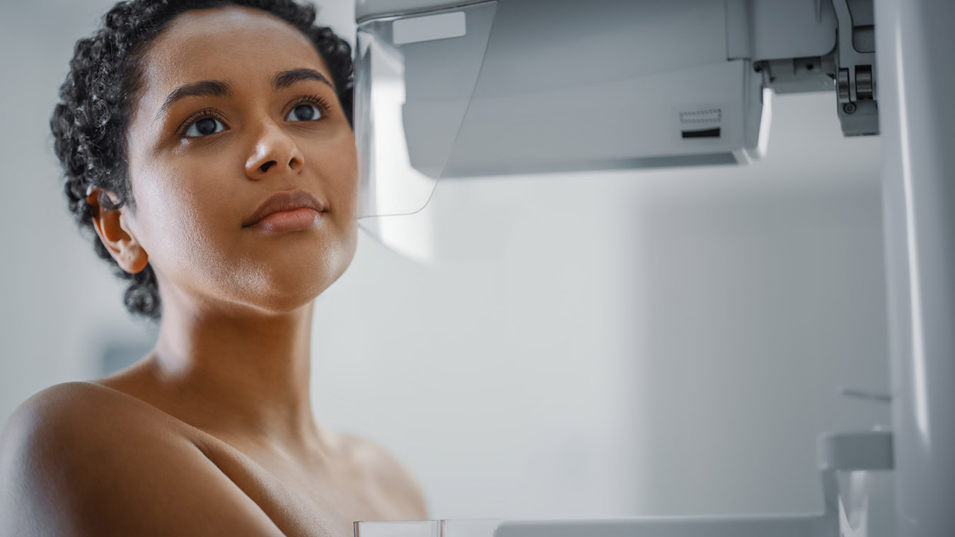
You will be asked to remove your clothing from the waist up and given a robe. Then your technologist will move you to the correct position between two X-ray plates. At this point, you will have the option to use a handheld remote to adjust the level of compression to what’s comfortable for you, under the supervision of the technologist. This will not affect the accuracy of the images and the technologist will ensure that the optimal level of compression is achieved for the clearest images. Or you can let the technologist control the level of compression.
The surfaces will slowly move together, gently compressing the breast for a short time to minimize your discomfort. Each breast will be imaged at least twice to create a top and side view for each one. It may be a bit uncomfortable, but it’s very quick, less than a minute of compression. The exam takes 10-15 minutes in total.
Once the pictures have been taken, one of our doctors will look over them very carefully to check for possible abnormalities or changes compared to previous images. This is the best way to detect breast cancer in its early, most treatable stage, because it provides a detailed look at the internal structure of breast tissue and can reveal changes that are too small to feel.
All women should have a mammogram. Many people start having regular mammograms every year at about age 40, since Alberta Health Care covers one mammogram per year starting at that age. If you have pain or a concern about your breasts earlier than that, you can of course go see your doctor and arrange to have a mammogram. To get a mammogram, you will need to speak to your doctor about your family history, when to start screening, and how frequently you should be screened.
Recommendations for breast screening intervals for women at regular risk:
Across Canada the recommended age to start screening and the recommended screening intervals differ by province. Mayfair’s recommendations for breast screening are aligned with the Canadian Association of Radiologists.
Women with the following risk factors are considered high risk and may be encouraged to start screening earlier and more frequently:
*Dense breast tissue refers to how it appears on the mammogram based on the mix of fatty and fibrous tissue. Women with very dense breasts may require a more personalized screening approach than what is recommended for the general population. This may include both mammography and ultrasound exams.
While the recommendations differ, the ultimate decision rests with women. Understanding the risks and benefits of regular mammogram screening and speaking with your doctor about your medical history is an important first step to decide what’s right for you.
Mayfair Diagnostics has 12 locations in Calgary and one in Cochrane which offer mammography exams. All of our locations except Coventry Hills feature patient-assisted compression. Visit our breast imaging page for more information.
REFERENCES
Alberta Health Services (2022) “Get Screened.” www.screeningforlife.ca. Accessed September 1, 2022.
Canadian Association of Radiologists (2016) “CAR Practice Guidelines and Technical Standards for Breast Imaging and Intervention.” www.car.ca. Accessed September 1, 2022.
Canadian Cancer Society (2022) “Breast cancer statistics.” www.cancer.ca. Accessed September 1, 2022.
Duffy, S.W., et al. (2020) “Mammography screening reduces rates of advanced and fatal breast cancers: Results in 549,091 women.” Cancer. Accessed September 1, 2022.
Monticciolo, Dr. et al. (2018) “Current Issues in the Overdiagnosis and Overtreatment of Breast Cancer.” American Journal of Roentgenology. February 2018, 210 (2). Accessed September 1, 2022.
Our Refresh newsletter delivers the latest medical news, expert insights, and practical tips straight to your inbox, empowering you with knowledge to enhance patient care and stay informed.
By subscribing to our newsletter you understand and accept that we may share your information with vendors or other third parties who perform services on our behalf. The personal information collected may be stored, processed, and transferred to a country or region outside of Quebec.
Please read our privacy policy for more details.
| Cookie | Duration | Description |
|---|---|---|
| cookielawinfo-checkbox-analytics | 11 months | This cookie is set by GDPR Cookie Consent plugin. The cookie is used to store the user consent for the cookies in the category "Analytics". |
| cookielawinfo-checkbox-functional | 11 months | The cookie is set by GDPR cookie consent to record the user consent for the cookies in the category "Functional". |
| cookielawinfo-checkbox-necessary | 11 months | This cookie is set by GDPR Cookie Consent plugin. The cookies is used to store the user consent for the cookies in the category "Necessary". |
| cookielawinfo-checkbox-others | 11 months | This cookie is set by GDPR Cookie Consent plugin. The cookie is used to store the user consent for the cookies in the category "Other. |
| cookielawinfo-checkbox-performance | 11 months | This cookie is set by GDPR Cookie Consent plugin. The cookie is used to store the user consent for the cookies in the category "Performance". |
| viewed_cookie_policy | 11 months | The cookie is set by the GDPR Cookie Consent plugin and is used to store whether or not user has consented to the use of cookies. It does not store any personal data. |